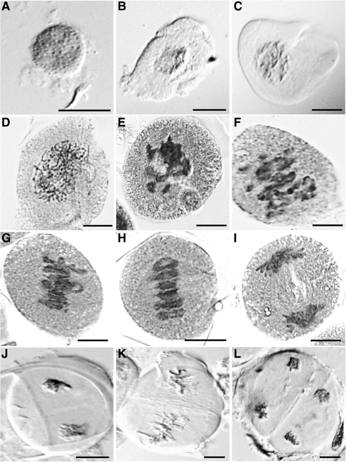
Fig. 5.
Light microscopic meiotic atlas of wheat showing the different stages of meiosis, a premeiotic G2 nuclei; b leptotene; c zygotene; d pachytene; e diplotene; f diakinesis; g early metaphase I; h metaphase I; i anaphase I; j telophase I; k metaphase II and (l) telophase II. The microscopic magnifications of the stages are different as the focus was to show chromosomal arrangements within the cells. Scale bar = 25 μm

