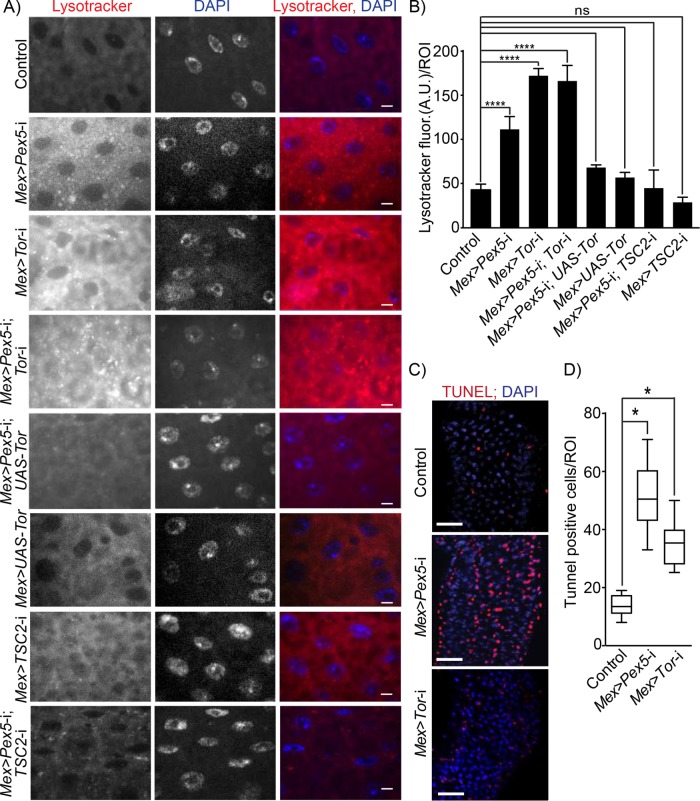
FIGURE 3:
Dysfunctional peroxisomes in the gut lead to increased Tor kinase-dependent autophagy and increased epithelial cell death. (A) Lysotracker staining (red) of midguts from flies of the designated genotypes. DNA was stained by DAPI (blue). Scale bar, 10 µm. (B) Quantification of Lysotracker fluorescence per ROI of midguts of flies of the designated genotypes. Values reported represent the averages of 20 independent ROIs ± SD for each genotype. Statistical significance was determined using two-way ANOVA; ****p < 0.0001; ns = not significant. (C) Midguts from Mex >Pex5-i and Mex >Tor-i flies exhibit increased numbers of apoptotic cells relative to midguts from control flies. Apoptotic cells were detected by TUNEL staining (red). Nuclei were stained by DAPI (blue). Scale bar, 25 μm. (D) Values reported represent the average number of TUNEL-positive cells per ROI per genotype for 25 midguts from each genotype. Statistical significance was determined using one-way ANOVA; *p < 0.05.

