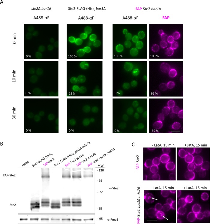
FIGURE 2:
Absence of yapsins preserves full-length endocytosis-competent FAP-Ste2. (A) Strain DK102 (ste2Δ bar1Δ) or otherwise isogenic derivatives expressing from the endogenous STE2prom, either Ste2-FLAG-(His)6 (yAEA265) or FAP-Ste2 (yAEA261), were incubated with A488-αF on ice for 1.5 h in medium lacking glucose and then washed and shifted to glucose-containing medium at 30°C, and samples were removed at the indicated times and viewed by fluorescence microscopy. The cells expressing FAP-Ste2 were prelabeled with fluorogen under standard conditions (0.4 mM dye; 15 min, 30°C, pH 6.5) prior to incubation with A488-αF. Value (%) in the bottom left corner of each image represents the average pixel intensity (n ≥ 200 cells per sample) of A488-αF or FAP-Ste2 at the cell periphery, relative to the starting intensity for each strain, quantified using CellProfiler, as described under Materials and Methods. Scale bar, 5 μm. (B) Strain JTY4470 (ste2∆) and otherwise isogenic yps1∆ or mkc7∆ single mutant derivatives or a yps1∆ mkc7∆ double mutant derivative (Table 1), expressing from the endogenous STE2prom either Ste2-FLAG-(His)6 or FAP-Ste2, as indicated, were grown to early exponential phase at 20°C, harvested, and lysed, and membrane proteins were extracted, resolved by SDS–PAGE, and analyzed by immunoblotting with anti-Ste2 antibody, as described under Materials and Methods. Loading control, Pma1 detected on the same immunoblots using anti-Pma1 antibody. MW, marker proteins (kDa). (C) Samples of a YPS1+ MKC7+ strain (yAEA152) or an otherwise isogenic yps1Δ mkc7Δ strain (yAEA359), each expressing FAP-Ste2, were treated, as indicated, with either vehicle alone (ethanol) or LatA in ethanol (100 μM final concentration) and then exposed to fluorogen as in A and viewed by fluorescence microscopy. Arrows, internalized vesicles containing FAP-Ste2. Scale bar, 5 μm.

