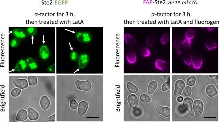
FIGURE 5:
Cells expressing FAP-Ste2 exhibit a normal morphological response to α-factor and insert newly made receptors at the shmoo tip. MATa cells expressing Ste2-EGFP (JTY6765) (left) and MATa yps1Δ mkc7Δ cells expressing FAP-Ste2 (yAEA359) (right) were treated with 10 μM α-factor for 3 h, incubated with LatA (and, in case of FAP-Ste2, then with fluorogen), and examined by fluorescence microscopy. Scale bar, 5 μm. Arrows, very slight enrichment of Ste2-GFP at shmoo tips (as compared with the prominent FAP-Ste2 fluoresence at shmoo tips).

