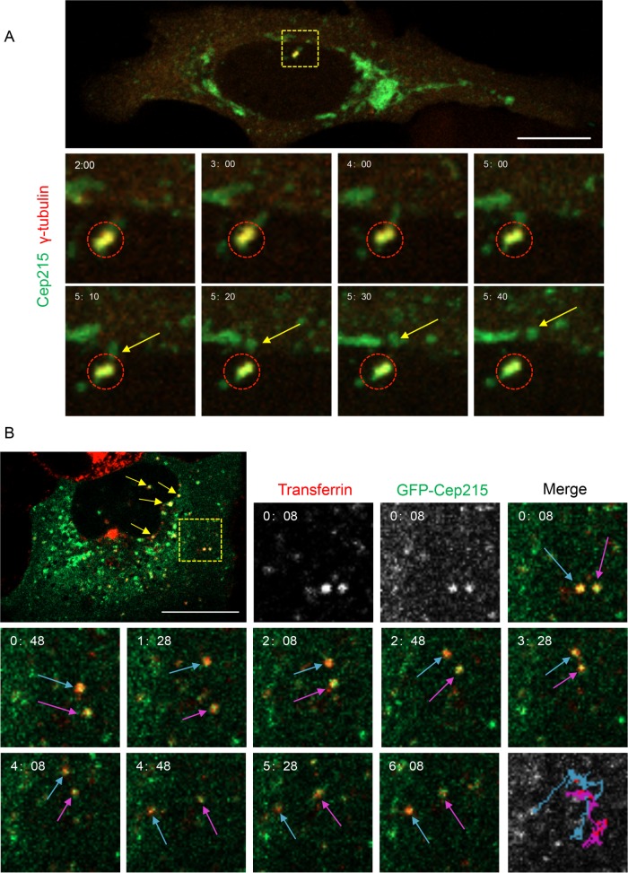
FIGURE 3:
Cep215 is transported away from centrosomes on transferrin-containing vesicles. (A) Cep215 displays directional movement away from the centrosome. U2OS cells transfected with pEGFP-Cep215 and dTomato-γ-tubulin for 16 h were imaged live every 2 s for 10 min. The dashed box indicates the γ-tubulin-labeled centrosome and is shown at higher magnification in the time-stamped insets. Circles mark the centrosome where Cep215 is localized. Arrows point to Cep215 moving away from the centrosome. Scale bar: 10 μm. (B) Cep215 localizes to mobile vesicles containing transferrin. U2OS cells transfected with pEGFP-Cep215 were pulsed for 15 min with Alexa Fluor 568–conjugated transferrin and imaged every 2 s for 10 min. Yellow arrows indicate Cep215 on transferrin-containing vesicles. Insets showcase two vesicles (dashed yellow box) at higher magnification. Blue and magenta arrows mark the movement of these vesicles, and their entire path is illustrated over time (bottom right panel). Videos compiled are representative of 18 (A) or 7 (B) time series obtained. Scale bar: 10 μm.

