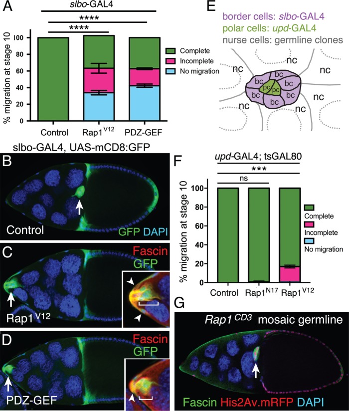
FIGURE 3:
Defined levels of activated Rap1 are required in specific cells for border cell migration. (A–D) Expression of constitutively activated Rap1, or elevated activation of Rap1 through PDZ-GEF, in border cells impairs border cell migration. (A) Quantification of complete (green), incomplete (pink), and no (blue) migration in stage 10 control, Rap1V12, and UAS-PDZ-GEF overexpression egg chambers. Genotypes: control (slbo-GAL4, UAS-mCD8:GFP/+), Rap1V12 (slbo-GAL4 UAS-mCD8:GFP/+; +/UAS-Rap1V12), PDZ-GEF (slbo-GAL4 UAS-mCD8:GFP/+; +/UAS-PDZ-GEF). Migration distance as in Figure 1B. Values consist of three trials, with each trial assaying n ≥ 100 egg chambers (total n ≥ 310 egg chambers per genotype); ****p < 0.0001; unpaired two-tailed t test, comparing “no migration.” Error bars: ± SEM. (B–D) Stage 10 control (B), Rap1V12 (C), and PDZ-GEF (D) overexpression egg chambers. slbo-GAL4 drives expression of UAS-Rap1V12 and UAS-PDZ-GEF, along with UAS-mCD8:GFP (green), in border cells (arrow), adjacent follicle cells, and centripetal cells (cells at the anterior side of the oocyte). DAPI (blue) labels nuclear DNA. Genotypes as in A. Insets, magnified view of the same border cell cluster costained with Fascin (red) to further label border cells (brackets) and adjacent follicle cells (arrowheads). (E) Schematic drawing of the border cell cluster, with the central polar cells, and surrounding nurse cells. Different GAL4 drivers can be used to test gene function in border cells (bc) and central polar cells (pc). Germline mosaic mutant clones can be used to test function in nurse cells (nc). (F) Rap1 function in polar cells. Quantification of migration at stage 10 when the polar cells express a control GFP (upd-GAL4/+; tsGAL80/UAS-PLC∆PH-GFP), Rap1N17 (upd-GAL4/+; +/UAS-Rap1N17; tsGAL80/+) or Rap1V12 (upd-GAL4/+; tsGAL80/UAS-Rap1V12), shown as complete (green), incomplete (pink), and no (blue) border cell migration. Migration distance as in Figure 1B. Values consist of three trials, with each trial assaying n ≥ 27 egg chambers per genotype (total n ≥ 134 per genotype); ns, p ≥ 0.05; ***p < 0.001; unpaired two-tailed t test comparing “complete” migration. Error bars in A and F: ± SEM. (G) Border cells complete their migration to the oocyte when nurse cells are mutant for a loss-of-function allele of Rap1. Representative example of a stage 10 Rap1CD3 mosaic mutant egg chamber stained for Fascin (green) to label the border cells (arrow) and DAPI (blue) to visualize nuclear DNA. His2Av.mRFP (red) marks the wild-type cells; loss of RFP marks homozygous mutant cells. In this egg chamber, all nurse cells are mutant (loss of red signal); the border cells and most follicle cells are wild type (colocalization of DAPI in blue and RFP in red appears as magenta).

