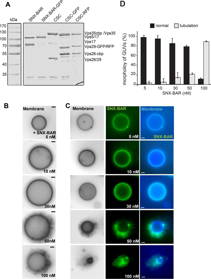
FIGURE 1:
Concentration-dependent membrane deformation by SNX-BAR proteins. (A) Purification of all proteins used in this study. Retromer subcomplexes with and without the indicated fluorescent tag were purified as described in Materials and Methods. (B, C) Membrane deformation by the Vps5-17 SNX-BAR dimer. The indicated amount of purified SNX-BAR complex without (B) or with (C) Vps17-GFP were titrated to GUVs at the indicated concentration in a 30-µl reaction volume, incubated for 15 min at room temperature, and then analyzed by fluorescence microscopy. Membranes were stained with Marina Blue DHPE lipid dye. Images of membranes were converted to black and white for better visualization. (D) Quantification of the tubulation by the tagged SNX-BAR complex. Data are represented as mean ± SD of three independent experiments. For details see Materials and Methods. Scale bar, 5 µm.

