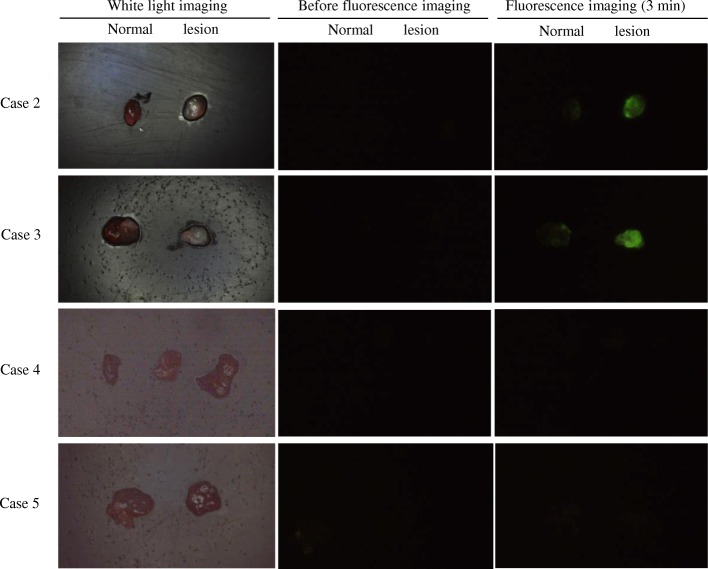
Fig. 3.
White-light and fluorescence images of PTC and non-neoplastic cases. In each figure, the left side represents normal tissue and the right side a lesion. Images were captured before and 3 min after gGlu-HMRG application. Case 2: PTC case. Green fluorescent light was observed. Case 3: PTC recurrence case. Recurrent papillary carcinoma also showed strong green signals from the gGlu-HMRG probe. Case 4: Adenomatous goiter case. In this case, almost the entire specimen was goiter tissue, and the three tissues were all goiter. The goiter tissue showed no green signal. Case 5: Chronic inflammation case. The lesion was suspected to be PTC but was thyroiditis. The thyroiditis showed no green signal

