
FIGURE 4:
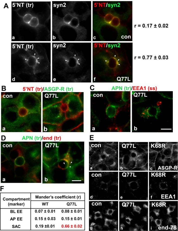
Transcytosing proteins accumulate in syntaxin 2–positive SAC structures in cells expressing GTP-bound/Q77L rab17. (A) Control (uninfected) WIF-B cells or cells expressing GTP-bound/Q77L rab17 were basolaterally labeled with antibodies against 5′NT and antigen-antibody complexes were chased for 60 min. Cells were fixed and double labeled for steady-state syntaxin 2 distributions. Merged images are shown in panels c and f Arrows indicate subapically accumulated transcytosing proteins in cells expressing mutant rab17. Bar = 10 μm. Mander’s coefficients of colocalization are indicated on the right. Values are expressed as the mean ± SEM from at least three independent experiments. Control (uninfected) WIF-B cells or cells expressing GTP-bound/Q77L rab17 were basolaterally labeled for 5′NT and ASGP-R (B) or APN (C) or APN and endolyn-78 (D) and allowed to continuously chase for 60 min. Cells were fixed and stained for the corresponding trafficked antibody–antigen complexes. In C, cells were labeled for steady-state distributions of EEA1. Merged images are shown for each. Arrows indicate subapically accumulated transcytosing proteins in cells expressing mutant rab17. Bar = 10 µm. In E, control (uninfected) WIF-B cells or cells expressing GTP-bound/Q77L or sumo-deficient/K68R rab17 were labeled for the steady-state distributions of ASGP-R, EEA1, and endolyn-78 as indicated. No changes in distributions were observed for any of the proteins confirming the validity of their use as compartment markers. Bar = 10 µm. In F, Mander’s coefficients of colocalization for the experiments shown in B, C, and D are shown. Values are expressed as the mean ± SEM from at least three independent experiments. BL EE, basolateral early endosome; AP EE, apical early endosome; SAC, subapical compartment.