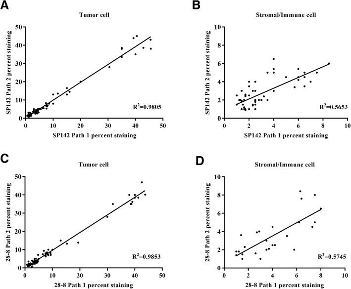
Fig. 4.
Inter-pathologist correlation of clone SP142 and clone 28–8 PD-L1 expression analysis. Scatter plot comparing the percentage of positive PD-L1 expression in tumor cell (a) and stromal/immune cell (b) from pathologist 1 and 2 using clone SP142. Scatter plot comparing the percentage of positive PD-L1 expression in tumor cell (c) and stromal/immune cell (d) from pathologist 1 and 2 using clone 28–8

