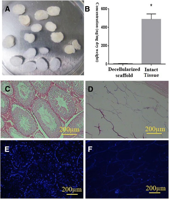
Fig. 1.
Macroscopic and microscopic structure of the rat testis after SDS-based decellularization process. A lyophilized decellularized testis scaffold showed whitish translucent appearance (a). The transverse section of the decellularized testis showed intact ECM and tunica albuginea gross architecture. DNA quantification showed significant cell removal by decellularization procedure (N = 3) (b). Comparing of the H&E (c, d), Hoechst (e, f), staining of the naïve (c, e), and decellularized tissue (d, f) also confirmed the cell removal. Both staining showed that ECM was preserved with no residual nuclei or cells

