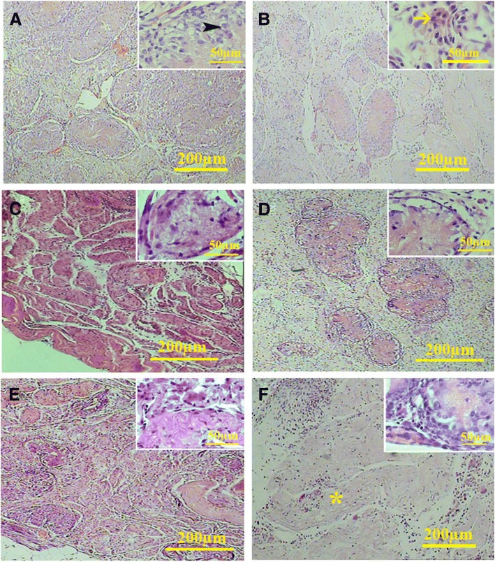
Fig. 11.
Histological sections of the cell-loaded (left series) and cell-free transplants (right series) on the mesentery. After 20 days (a, b), the cells within the seminiferous tubule showed sertoli-like cell phenotype (arrow head) along with round cells with leydig cell-like phenotype in the interstitium (arrow). A similar phenotype could be observed after 40 days (c, d). After 60 days, some tubules contained sertoli-like cells that had yet occupied some tubules. The percentage of the tubules occupied by fibrous tissue increased significantly at day 60 (asterisk) (e and f)

