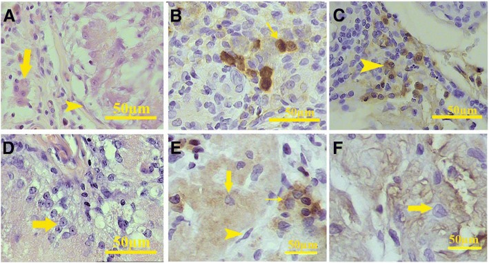
Fig. 12.
Higher magnification of the H&E staining showed that some cells with eosinophilic cytoplasm and round nuclei were present in the interstitium (arrow) after 20 days. Also, some elongated cells with close similarity with myoid cells present in the tissue surrounded each seminiferous tubule (arrowhead, a). Immunohistochemistry showed that some cells within the interstitium reacted with anti-inhibin antibody (b). Immunohistochemistry for CD68 revealed that a subpopulation of the round cells located between the tubules were macrophage (arrowhead, c). Also, H&E staining shows the presence of the cells with pale trigonal or round nuclei and expanded cytoplasm in some tubules in all samples (arrow, d). These sertoli-like cells were present within the tubules and react with anti-inhibin antibody (large arrow). Leydig-like cells that express inhibin (small arrow) and myoid-like cells (arrowhead) also were present in the transplanted scaffold (e). After 60 days, some seminiferous tubules contain inhibin expressing sertoli-like cells (arrow, f)

