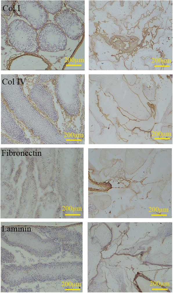
Fig. 4.

Comparison of the ECM content of the intact testis (left series) with decellularized scaffolds (right series) showed that collagen I, collagen IV, fibronectin, and laminin remained after decellularization. The distribution of these proteins was the same in both cellularized and decellularized tissues. The laminin and collagen IV were mainly present in the basal lamina, while the fibronectin distributes in all regions uniformly
