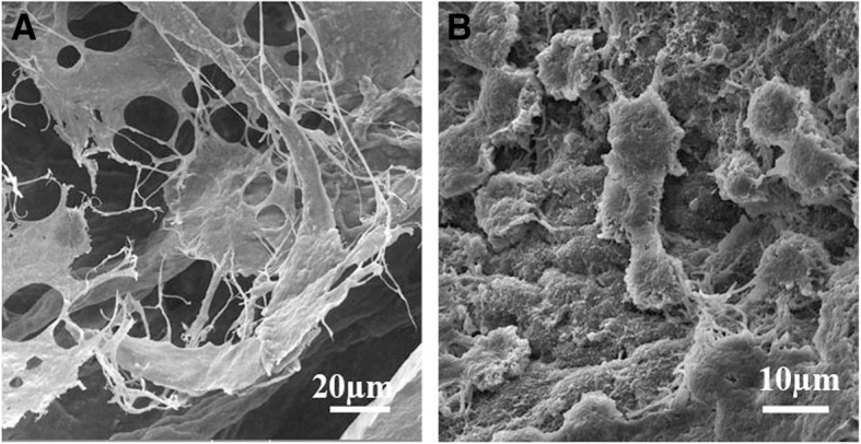
Fig. 6.

Scanning electron microscopy of the different regions of the recellularized scaffolds. Electron micrographs revealed that the cells were attached to the scaffolds; the cells located within the interstitium showed a cell body with numerous processes (a), while those located within the seminiferous tubules were spherical (b)
