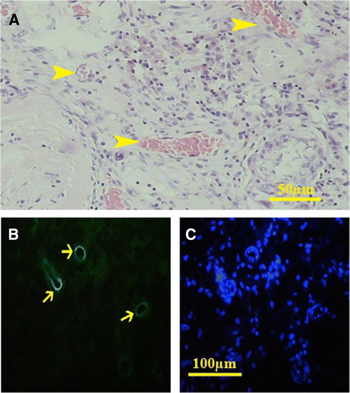
Fig. 9.
In vivo study showed that angiogenesis happened properly and different sections of the blood vessels were present in the transplanted scaffolds (a). H&E staining showed that all types of vessels were present in the transplanted sections. UEA lectin, as a marker of endothelial cells, also confirmed the angiogenesis (b, FITC-conjugated UEA; c, Hoechst)

