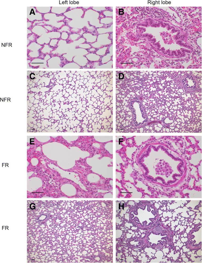
Fig. 5.

Representative histology showing endotoxemia and saline resuscitation associated changes to bronchial and alveolar histology. The left lobe from NFR (a, c) and FR (e, g) groups had minimal inflammatory infiltrate or oedema. Right lobe tissue in NFR (b, d) and FR (f, h) groups had some additional damage to bronchioles. FR = fluid resuscitation; NFR = no fluid resuscitation. Scale bars = 50 μm
