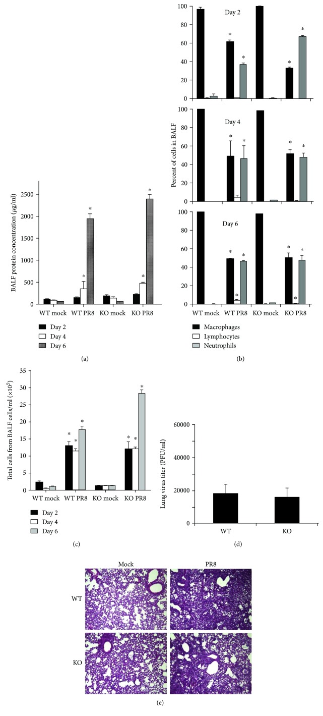
Figure 2.
Inflammatory profile in the bronchoalveolar lavage fluid (BALF) and virus titer in the lung. WT and RIG-I KO mice were intranasally infected with 300 pfu of the IAV PR8 or mock infected with PBS. BALF was harvested at the indicated time points after infection. Total protein levels (a), immune cell differential (b), and total cells (c) in BALF were determined. Cytospins of the cells were prepared using a Cytopro Cytocentrifuge and stained with Diff-Quik. Differential counts were manually determined using the morphology of more than 400 cells/sample from 2 slides/mouse. Lung tissue viral titers were determined at 6 days postinfection by plaque assay on MDCK cells (d). Data are expressed as means ± SEM (n ≥ 3/group). ∗ denotes significant difference compared to the corresponding mock infected groups (p < 0.05). (e) Mouse lung tissue pathology after IAV infection. Mice were intranasally infected with 300 pfu of IAV PR8 strain. Samples were harvested after 6 days. Lung tissue sections prepared from the infected mice were fixed, processed, and stained with H&E. The lungs of 3 mice from each treatment group were processed for histology, and results shown were typical for the group (200x magnification).

