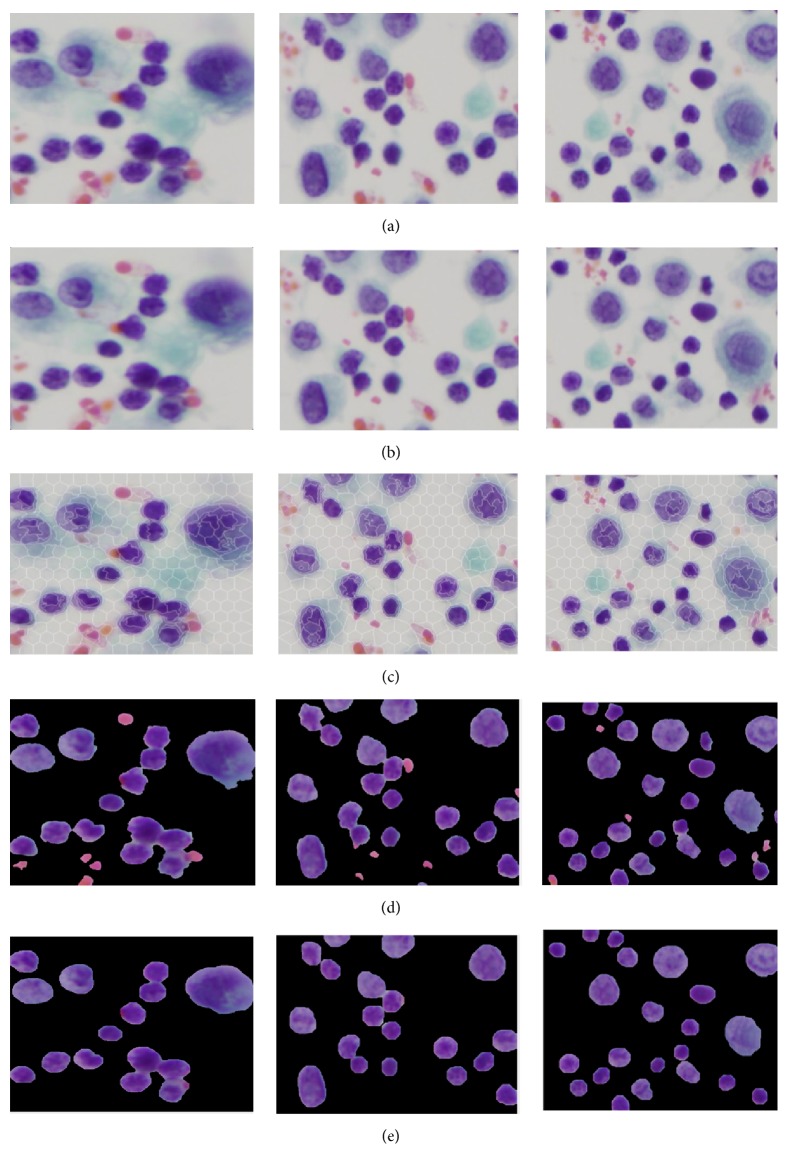
Figure 3.
Visual results of segmenting cell nuclei from CPE images: (a) original image, (b) preprocessed image, (c) superpixels segmentation using SLIC, (d) K-Means based unsupervised color segmentation on SLIC superpixels, and (e) postprocessed image (refinement of nuclei boundary and elimination of false findings).

