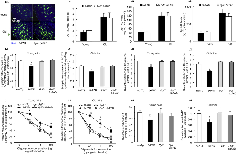
Figure 2. CypD deficiency attenuates mitochondrial F1Fo ATP synthase deregulation in 5×FAD mice.
(a) Aβ levels in 5×FAD and CypD deficient 5×FAD mice in young and old age. n=6 for each group. Scale bar, 100μm. ELISA from isolated synaptic mitochondria showed similar levels of Aβ 1–40 and Aβ 1–42 in both AD genotype mice. (b) Synaptic mitochondria from 5×FAD mice demonstrated blunted F1Fo ATP synthase catalytic activity, which exacerbates with age. This defect was protected by CypD deficiency in both young and old Ppif−/− 5×FAD mice. n=5–7 per group. *P<0.05 vs other groups. (c) Decreased oligomycin sensitivity of synaptic mitochondria from young and old 5×FAD mice were rescued by CypD deficiency. All data are presented as percentage of the activity of the corresponding vehicle-treated mitochondrial fractions. n=5–7 per group. *P<0.05 vs other groups. (d) Synaptic mitochondria from 5×FAD mice showed an age-dependent decrease in RCR, which was protected by CypD depletion. n=5–7 mice per group. *P<0.05 vs other groups (e) Synaptic mitochondria from 5×FAD mice demonstrated an age dependent decline in ATP synthesis as compared to other groups. n=5–7 mice per group. *P<0.05 vs other groups. Error bars represent s.e.m.

