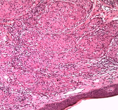
Figure 4.

Histology result of the patient’s biopsy. Epithelioid granulomas with lymphocytes present on the periphery. Caseation necrosis absent. The vessels of the superficial vascular plexus have a dilating lumen. Disseminated, distinct, non-caseating naked granulomas visible.
