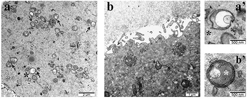
Figure 3.

Representative TEM images of a) parthenogenetic NSN-derived 2-cell embryo with vesicular mitochondria (black arrows and black asterisk); b) SN-derived 2-cell embryo obtained by ICSI showing non-defective mitochondria (white asterisk); a’ and b’ magnification of a vesicular and normal mitochondria respectively.
