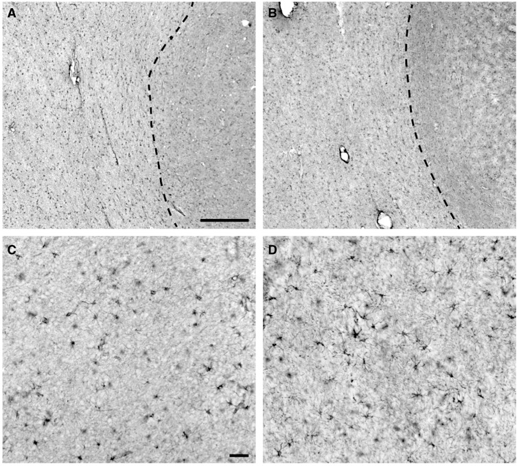
FIGURE 3.
Immunohistochemical visualization of glial fibrillary acidic protein (GFAP) labeling in prefrontal white matter samples with and without vascular brain injury (VBI). (A) Representative GFAP labeling of astrocytes in a No VBI case and a VBI case (B) , both taken at low power, demonstrate the homogenous distribution of GFAP-positive cells within the underlying white matter. Dashed line demarcates the cerebral gray/white matter borders. (C) A higher power image of GFAP labeling in the white matter in the same No VBI case as in (A) . Note the presence of heavy GFAP labeling of the ramified processes of many of the astrocytes. Overall, the labeled processes are indicative of normal astrocyte morphology. (D) GFAP labeling of astrocytes in the same VBI case as shown in (B) . Morphology of the cells is representative of astrocytes, but a slight increase in proximal process thickness and ramification of more distal processes indicates that these likely are reactive. The subtle visual differences between these panels underscore the challenge of differentiating between moderately reactive and nonreactive astroglial phenotypes when evaluated by low-power, 2-dimensional fields or through similar conventional histopathologic methods. Scale bar: A = 500 µm and also applies to B; C = 50 µm and also applies to D.

