Fig. 4.

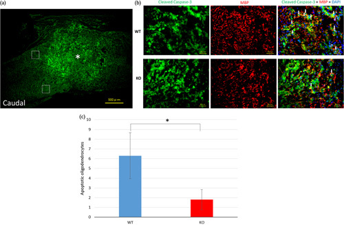
Spinal cord sections from day 7 after spinal cord injury (SCI) immunostained for cleaved caspase-3, myelin basic protein (MBP), and 4′,6-diamidino-2-phenylindole dihydrochloride (DAPI). (a) The asterisk shows the epicenter of the injury site (scale bar=500 µm). The white squares indicate the two regions in the white matter. (b) Enlarged images of the regions (scale bar=40 µm) are shown. These digital images show apoptotic cells immunostained for cleaved caspase-3 (green) and oligodendrocytes stained for myelin basic protein (MBP; red) and DAPI (blue). The white arrows show trichrome-stained apoptotic oligodendrocytes. (c) The cells co-labeled with cleaved caspase-3 and MBP were defined as apoptotic oligodendrocytes. The number of apoptotic oligodendrocytes immunostained for cleaved caspase-3, MBP, and DAPI at the injury site. The number of trichrome-stained cells was significantly higher in the wild-type (WT) mice than in the knockout (KO) mice (*P<0.05, Student’s t-test).
