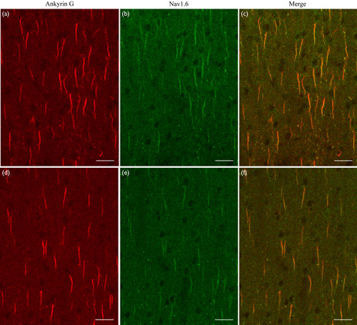
Fig. 3.

Immunofluorescent double labeling showing the axon initial segment (AIS) (red) and the distribution of Nav1.6 Na+ ion channels (green) along AISs of layer II/III neurons in the V1 cortical area of young adult (a–c) and aged (d–f) rats. (a, d) AISs of neurons labeled with anti-AnkyrinG, a specific scaffolding protein. (b, e) The distribution of Nav1.6 Na+ ion channels along AISs. (c, f) Merged images of AnkyrinG and Nav1.6 fluorescence along AISs. The scale bar represents 30 μm.
