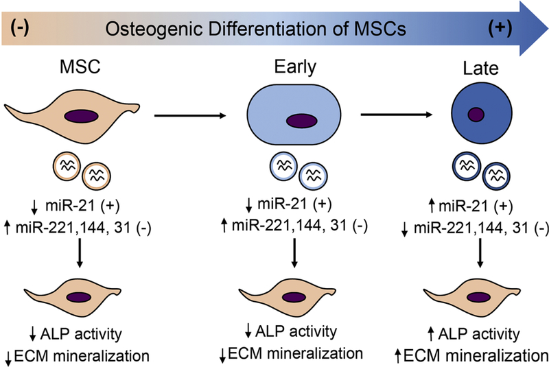
Figure 4. Schematic describing that EVs from varying stages of differentiating MSCs induce osteogenic differentiation of MSCs differently.

Osteogenic differentiation was induced in MSCs using osteogenic differentiation media. EVs were isolated from conditioned media collected at different stages after media change, namely, early and late stages. Exosomes from MSCs in growth media were used as control. Using an array-based method, expression of miRNA associated with osteogenic differentiation of MSCs was measured. ALP activity, ECM calcium, and phosphate was measured to evaluate osteogenic differentiation of MSCs after EV treatment. Adapted from data presented in Wang et al., (2018) via open access.
