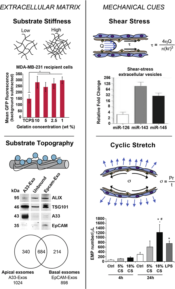
Figure 5. ECM characteristics and mechanical stimulation of cells can dictate EV production, composition, and uptake profiles.

In the top left figure, breast cancer cell lines MCF-7 and MDA-MB-231 were seeded on gelatin substrate with varying stiffness. MDA-MB-231 cells seeded on gelatin matrix with different stiffness were treated with CD63-GFP-labled EVs. EV uptake was monitored following 2 hours of treatment through live-cell imaging. Seeding cell on softer gelatin matrices compared to tissue culture polystyrene (TCPS), resulted in significantly increased EV uptake for both cell lines. Adapted from Tauro et al., (2013) via open access. Bottom left figure shows western blot analysis of exosomes (10 µg) derived from LIM1863 cells from apical (EpCAM-Exos) or basolateral (A33-Exos) surfaces, which were enriched in EpCAM and A33, respectively. Both sources were also positive for exosomal markers TSG101 and Alix. Venn diagram shows differences in protein expression between EpCAM-Exos and A33-Exos. In total, 340 and 214 proteins were specifically found in A33-and EpCAM-Exos, respectively. In comparison, 684 proteins were present in both exosome sources. Adapted from Stranford et al., (2017) with permission. For the top right figure, EVs were isolated from HUVECs with or without exposure to shear-stress of 20 dynes/cm2 for 3 days. RNA analysis revealed that EVs derived from HUVECs under shear stress were highly enriched in miR-143/145, which are known to modulate SMC phenotype. Adapted from Hergenreider et al., (2012) with permission. For the bottom right figure, confluent lung-ECs were subjected to 5% CS or 18% CS for 4 and 24 hours or to LPS (1 µg/mL) for 24 hours. Lung-EC-derived microparticles (EMPs) (0.1–1 µm) were isolated from conditioned media and quantified by flow cytometry. Significant increase in EMP production is observed after 24 h exposure to 18% CS and LPS compared to the static control. Reprinted from Letsiou et al., (2015) with permission.
