
Figure 1: The basement membrane is damaged by pinch wounds and forms a scar upon repair.
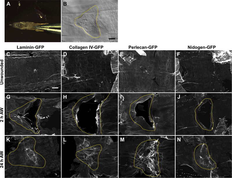
A) To damage the basement membrane, blunt forceps were used to pinch larvae on the dorsal epidermis between the hair stripes of segments A3 and A4. B) Pinch wounds do not break the outer cuticle, but they do leave an indentation visible with DIC, outlined with yellow dotted line. C-F) Undamaged epidermal basement membrane visualized with GFP-fusion constructs of each of the core basement membrane proteins. Images are representative of the no-knockdown controls quantified in Fig. 3. G-J) Damaged epidermal basement membrane after pinch-wounding. K-N) Within 24 h, the basement membrane was repaired, leaving behind a visible scar in the region of the healed wound. Images are representative of the no-knockdown controls quantified in Fig. 4. Dotted yellow lines indicate original wound borders based on cuticle indentation. Scale bar, 50 µm.