
Figure 2: The basement membrane scar is thicker than unwounded basement membrane.
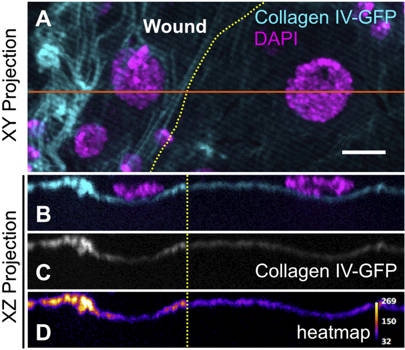
Yellow dotted lines indicate wound border. Orange solid line indicates location sampled for XZ projections. A) Basement membrane scar, evident on the left (wounded) side. B-D) Z-section shows increased thickness and fluorescence of collagen IV within the healed wound (N ≥ 3). Scale bar, 10µm.