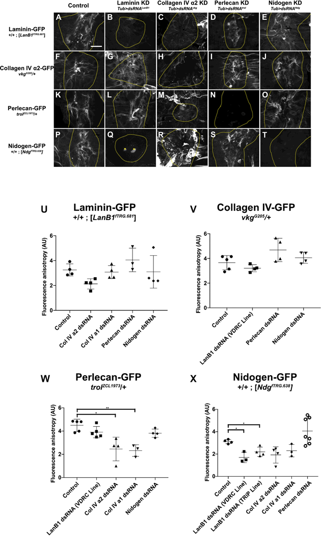
Figure 6: Hierarchy of basement membrane assembly during repair.

A-E) Laminin assembled into repaired basement membrane independent of any other basement membrane proteins. F-J) Collagen IV assembled into repaired basement membrane independent of any other basement membrane proteins. K-O) Although perlecan assembled into repaired basement membrane independent of any other basement membrane proteins, its assembly into the scar required collagen IV (M). P-T) Nidogen required laminin (Q) but not collagen IV (R) or perlecan (S) to assemble into repaired basement membrane. In collagen knock-down wounds (C,H,M,R), scars appear to extend outside the wound area, see text. In panel Q, the bright dots at the wound center (marked by yellow stars) are autofluorescent melanization, see Experimental Procedures. U-X) Quantification of fluorescence anisotropy in repaired basement membranes. * indicates p ≤ 0.05, ** indicates p ≤ 0.01. Unless otherwise indicated, no significant difference was observed. Scale bar, 50 µm.
