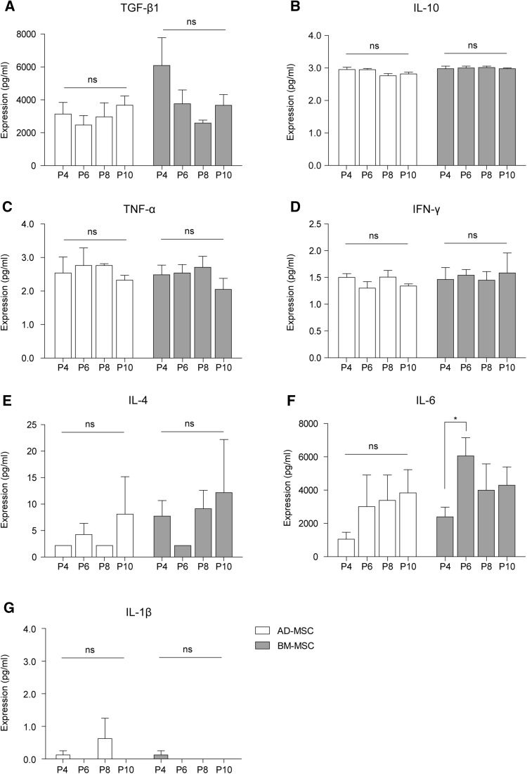
Fig. 2.
Human AD- and BM-MSCs cytokine expression through passages. A The levels of TGF-β1 were higher in BM-MSC P4 than late passage. But, this was not significantly different in AD- and BM-MSCs through passages. B–E, G Human IL-10, TNF-α, IFN-γ, IL-4, and IL-1β were scantly produced from P4, 6, 8, and 10 by AD- and BM-MSCs. F IL-6 was highly expressed in both MSC types. This was significantly higher in BM-MSC P6 compared to P4, however there was no significant difference between the other passages. Experiments were performed in triplicate for each sample. All values are the mean ± SEM. *p < 0.05 was considered statistically significant versus P4 of each origin, ns means not significant

