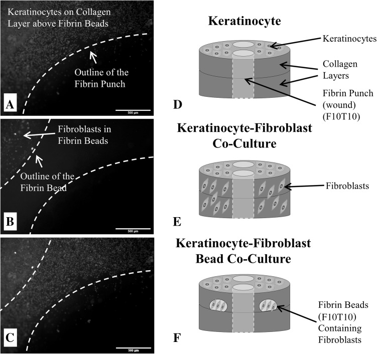
Fig. 1.
Micrographs of A keratinocytes fluorescently-tagged with Vybrant DiD on the surface of the 3D wound healing construct and near the fibrin-filled defect at a concentration of 300,000 cells/mL; B fibroblasts fluorescently-tagged with Vybrant DiO that are encapsulated inside a fibrin bead within the collagen layers of the 3D wound healing construct and near the fibrin-filled defect (20,000 cells/bead); and C a stacked fluorescent image of the keratinocyte-fibroblast bead co-culture with fibroblasts encapsulated inside fibrin beads. For all micrographs, the scale bar = 500 µm (50 x magnification). D Illustration of the control construct with only keratinocytes seeded on the surface. E Illustration of the keratinocyte-fibroblast co-culture construct with fibroblasts uniformly dispersed throughout the collagen matrix and keratinocytes seeded on the surface. F Illustration of the keratinocyte-fibroblast bead co-culture with fibroblasts encapsulated inside two fibrin beads within the collagen matrix and keratinocytes seeded on the construct’s surface. The final fibrin concentrations were either 5 or 10 mg/mL (F5 or F10) and polymerized with either 5 or 10 units/mL thrombin (T5 or T10). The concentration of the fibrin-filled defect matched the fibrin bead formulation. Note the illustrations are not drawn to scale

