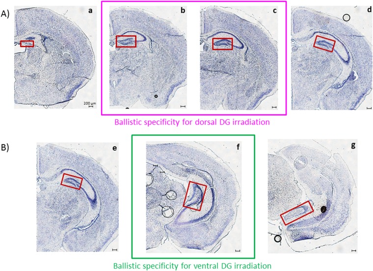
Figure 3.
Distinct transverse sections observed for ballistic validation. Cresyl violet staining has been used here to reveal nervous tissue structures. The DG is framed in red in the photomicrographs. Four and three transverse sections were selected for the validation of dorsal (A) and ventral (B) DG model of irradiation, respectively. Sections a, d, e and f are supposed to be in the non-irradiated field, whereas sections b, c and f are supposed to be in the irradiated zones when following targeted irradiation protocols.

