Figure 1.
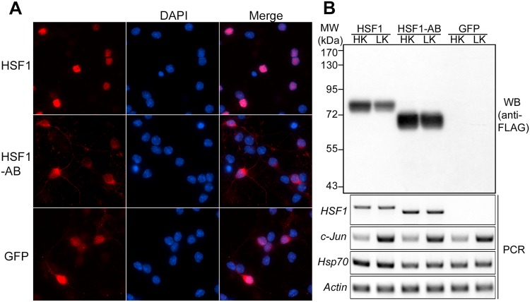
Overexpression of HSF1 and HSF1-AB in CGNs. (A) CGNs were infected with Ad-HSF1, Ad-HSF1-AB, or Ad-GFP and treated with HK or LK for 8 hours. Immunostaining using FLAG or GFP antibody showed that HSF1 was mainly localized in the nucleus, while HSF1-AB and GFP were distributed in both the cytoplasm and the nucleus. (B) HSF1 and HSF1-AB were robustly expressed in the CGNs as shown by Western blot (WB) and RT-PCR analyses. Expression level of Hsp70, a known target gene of trimeric HSF1, was upregulated by wild-type HSF1 but not by HSF1-AB, which lacks the trimerizaiton domain. Level of an apoptotic marker, c-Jun, was dramatically increased by LK treatment. The darker intensity of signal for HSF1-AB relative to HSF1 is because of its higher stability (Qu and D’Mello, manuscript in preparation).

