
Figure 2.
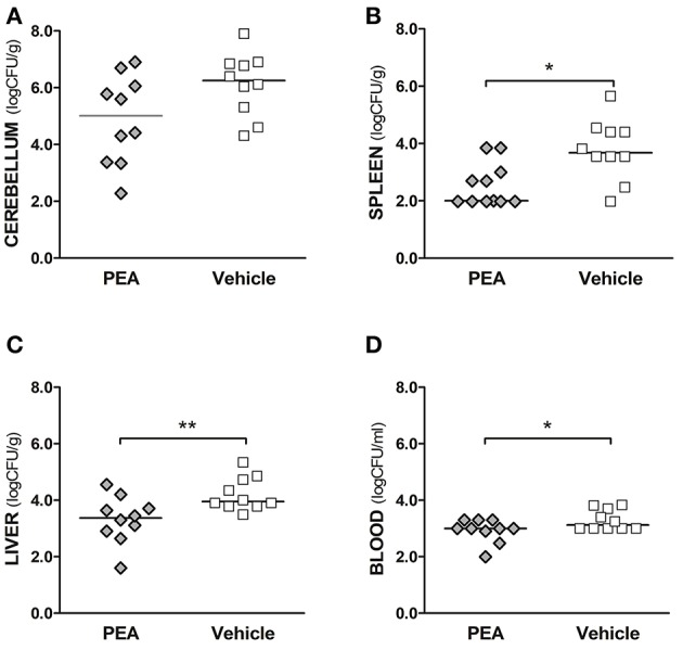
Exogenous PEA reduces bacterial spread in aged mice after the induction of E. coli K1 meningoencephalitis. Concentrations of E. coli K1 were quantified in (A) cerebellum, (B) spleen, and (C) liver (as log10CFU/g), as well as in (D) blood (as log10CFU/ml) of animals sacrificed 24 h after infection with 3,000 CFU E. coli K1/mouse (n= 10 mice/group, data from two independent experiments). Each symbol represents an individual mouse and bars indicate median values. **P < 0.01, *P < 0.05 between PEA and vehicle groups, using the Mann–Whitney U-test.