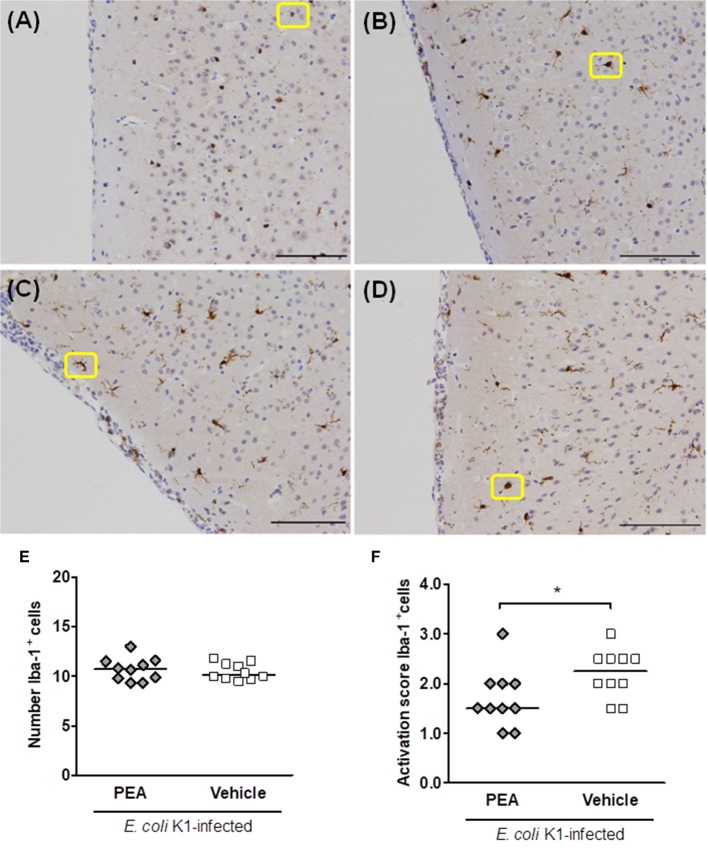
Figure 5.
PEA prophylaxis does not modify microglial density, but attenuates microglial activation in aged infected mice. (A–D) Illustrative examples of the different cell morphologies (circled in yellow) and the corresponding activation scores in neocortex. A single activation score (AS) was given by a blinded investigator according to the most abundant cell morphology for each analyzed region: (A) microglia with small size and very fine ramifications (AS 1), (B) hypertrophic with thicker branches (AS 2), (C) bushy (AS 3), and (D) ameboid (AS 4); scale bars, 100 μm (magnification, ×20). (E) Number of Iba-1+ cells in brains of PEA- and vehicle-treated aged mice sacrificed 24 h after infection. (F) AS of Iba-1+ cells in brains from the same PEA- and vehicle-treated aged mice. In (E,F), each symbol represents an individual mouse and bars indicate medians. *P < 0.05, using the Mann–Whitney U-test.

