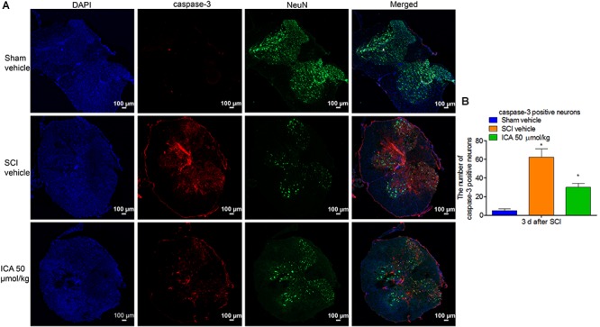
FIGURE 5.

Immunofluorescence staining of caspase-3/NeuN/DAPI. (A) Immunofluorescence staining (Scale bars = 100 μm). (B) The number of caspase-3+ neurons was dramatically increased after SCI. However, 50 μmol/kg ICA significantly decreased caspase-3 positive neurons. N = 6. ∗P< 0.01 compared to SCI group.
