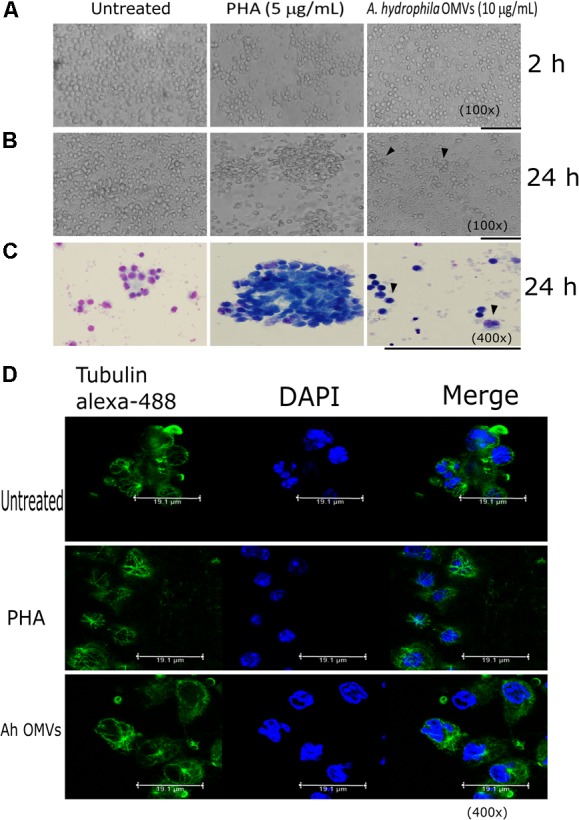
FIGURE 7.

Morphological alterations induced by A. hydrophila ATCC® 7966TM OMVs in PBMCs. (A) After 2 h post-stimulation with A. hydrophila OMVs, no morphology changes of the PBMCs were observed compared with PBMCs after 24 h post-stimulation with vesicles, after 24 h post-stimulation conglomerated cells were observed (arrowheads) (B). Giemsa staining revealed differences in the affinity of the dye to the nucleus and cytoplasm, bigger cells, and vacuolization of the stimulated PBMCs with A. hydrophila OMVs (arrowheads) (C). Confocal microscopy, showed defective tubulin polymerization in PBMCs stimulated with OMVs (D). bars = 50 μm.
