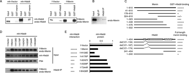
Figure 2.
Hlxb9 interacts with menin. (A) GST pull-down assay. WCE from MIN6 cells transfected with mychis-Vector (mh-Vector) or with mychis-Hlxb9 (mh-Hlxb9) plasmid (left panel) was incubated with GST or GST-menin beads. Bound Hlxb9 was detected by western blot. Input (1/20th amount of WCE incubated with beads) was analyzed side-by-side. WCEs of cells transfected with flag-Vector (f-Vector) or flag-Menin (f-Menin) plasmid (right panel) were incubated with GST or GST-Hlxb9 beads. Bound menin was detected by western blot. Myc-Hlxb9 and endo-Hlxb9 indicate transfected mychis-Hlxb9 protein and endogenous-Hlxb9 respectively. F-Menin and endo-Menin indicate transfected flag-menin protein and endogenous menin respectively. (B) Co-immunoprecipitation assay. WCE immunoprecipitated with IgG or anti-Hlxb9 followed by western blot with anti-menin. Input lane shows 1/20th amount of WCE used for immuno-precipitation. (C) Menin:Hlxb9 interaction regions. Top panel shows a schematic diagram of full-length menin (aa 1–610) and menin deletion constructs containing the indicated amino acids from the N- or C-terminal region. Bottom panel shows a diagram of GST-fused full-length Hlxb9(aa 1–404) and GST-fused Hlxb9 constructs with the indicated internal deletions or containing the indicated amino acids from the N- or C-terminal region. In Hlxb9 (bottom panel), the left slanting hatched boxes indicate poly-alanine containing regions; right slanting hatched box indicates conserved domain, and crosshatched box indicates the homeodomain. The binding ability of menin constructs to GST-Hlxb9 or to GST-Hlxb9 deletions is represented by ‘+’ sign, ‘+’ is lowest binding, ‘+++++’ is highest binding. Western blot images from GST-Hlxb9 pull-down assays are shown in Supplementary Figure 3B and C. (D) Hlxb9 interaction with menin missense mutants. WCEs of MIN6 cells expressing mh-Hlxb9 together with f-Menin or indicated f-Menin missense mutants were assessedfor Hlxb9–menin interaction by immunoprecipitation with anti-Hlxb9 followed by western blot with anti-menin. Menin (normal and missense mutants) and Hlxb9 levels are shown in the western blots in the upper panels (input). P84 was used to assess protein loading. f-Menin and endo-Menin corresponds to transfected flag-Menin and endogenous menin respectively. (E) Hlxb9 interaction and expression. Data corresponding to Hlxb9 interaction with menin and Hlxb9 expression from the western blots in (D) are shown. The interaction ability of Hlxb9 is represented by ‘+’ sign, ‘+’ is lowest binding, ‘+++++’ is highest binding.

