
Figure 2.
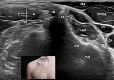
Roots/rami of the brachial plexus – axial view. A: anterior. AT: anterior tubercle. CA: carotid artery. CVB: cervical vertebral body. IJV: internal jugular vein. L: lateral. LCM: longus colli muscle. M: medial. P: posterior. PED: pedicle. PM: platisma muscle. PT: posterior tubercle. SCMM: scalenus medium and posterior muscle. SR: spinal root. VN: vagus nerve. The probe position is indicated by the white bar in the insert.