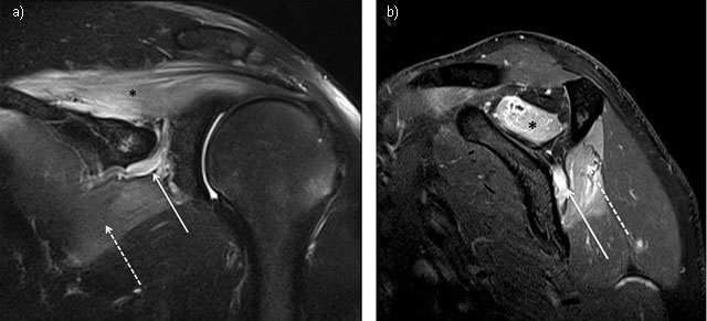
Figure 1.

Suprascapular neuropathy at the scapular notch in a young judoka athlete showing typical edema denervation pattern of acute neuropathy on MRI. High signal intensity fluid is observed in fluid-sensitive sequences of both supraspinatus (star) and infraspinatus (dashed arrow). Notice the dilatation of suprascapular veins satellite (arrow). (1a = FIGURE 1 uploaded online manuscript) – Coronal (1b = FIGURE 2 uploaded online manuscript) – sagittal Short Tau Inversion recovery MRI (STIR) images.
