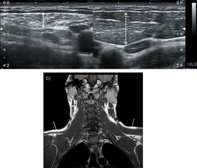
Figure 5.

Chronic neuropathy of spinal accessory nerve. Ultrasound showed atrophy of sternocleidomastoid muscle (double solid arrow) compared to normal side (doubled dashed arrow) (5a = FIGURE 13 uploaded online manuscript), confirmed by coronal T1-weighted MRI showing both atrophy of trapezus (solid arrow) and sternocleidomastoid muscles compared to normal side (dashed arrow) (5b = FIGURE 14 uploaded online manuscript).
