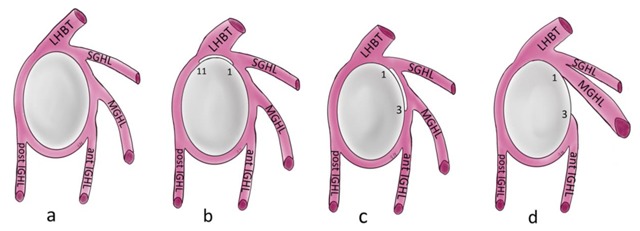
Figure 12.

Schematic illustration of the normal capsulolabral complex and anatomical variations. They are shown on a lateral view onto the glenoid. (a) Normal anatomy; (b) Sublabral recess (sublabral sulcus); (c) Sublabral foramen (sublabral hole); (d) Buford complex. LHBT: long head of biceps tendon, SGHL: superior glenohumeral ligament, MGHL: middle glenohumeral ligament, IGHL: inferior glenohumeral ligament.
