


Figure 13.
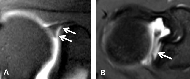
Sublabral recess (sublabral sulcus). (A) Coronal oblique fat-suppressed T1-weighted MR arthrographic image shows a sublabral recess as an increased linear signal undercutting the contour of the superior glenoid labrum (arrows, A) following the contour of the glenoid cartilage without extension posterior to the biceps anchor. This sulcus is visualized on (B) Axial T1-weighted MR arthrographic image (arrow, B) on an upper section.