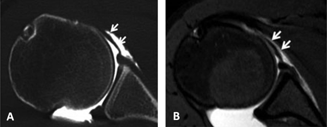
Figure 20.

Middle glenohumeral ligament. The midsection of the ligament often adopts a more horizontal course. This can result in a thinner, wavier appearance on axial fat-saturated proton density MRA image (arrows, B) with a longer section of the ligament (arrows) on CTA (A).
