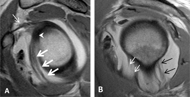
Figure 23.

Inferior glenohumeral ligament. (A) Sagittal oblique PD-weighted MRA depicts the inferior glenohumeral ligament (thick arrows, A) with a high labral attachment (arrowhead, A). Coracohumeral ligament (thin arrow, A). (B) Sagittal oblique PD-weighted MRA shows the anterior band of the inferior glenohumeral ligament (white arrows, B) and the posterior band of this ligament (black arrows, B).
