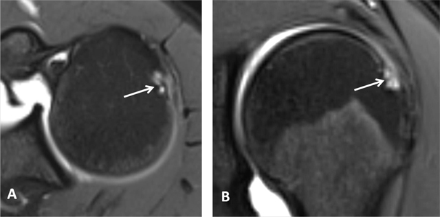
Figure 3.

Subchondral cysts of the humeral head and normal bare area. (A) Axial and (B) Coronal oblique fat-suppressed T1-weighted MR arthrographic images show subchondral cysts at the attachment of the infraspinatus tendon (arrow). Coronal oblique section of the same patient discloses a normal bare area in the posterolateral aspect of the humeral head with small fibrocystic changes that communicate with the joint and should not be mistaken for a cartilage defect (arrow, B). Such changes are common and often asymptomatic.
