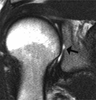
Figure 6.

Bare area of the glenoid on MRI. Coronal oblique PD-weighted MR image displays a defect in the cartilage filling up with a moderate amount of joint fluid (arrow) without any thickening of the subchondral bone. As for the tubercle of Assaki, the bare area of the glenoid may be mistaken for a cartilage ulceration.
