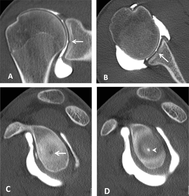
Figure 7.

Bare area of the glenoid on CTA. (A) Coronal, (B) Axial and (C, D) Sagittal reconstructed CTA images demonstrate in the mid third of the glenoid a defect of the hyaline cartilage located centrally (arrows, A–C). On D, the defect is filled up by the injected contrast (arrowhead).
