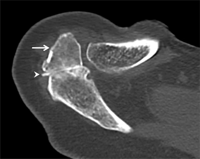
Figure 9.

Os acromiale. Axial CT arthrography through the acromioclavicular joint demonstrates an os acromiale (arrow) with synchondrosis (arrowhead). An os acromiale should not be confused with a fracture fragment and the synchondrosis with a pseudarthrosis. In this case, there is a communication between the synchondrosis and the glenohumeral joint (with high-density contrast penetration), which is not normal. Subchondral cystic changes are also seen in the zone of pseudarthrosis.
